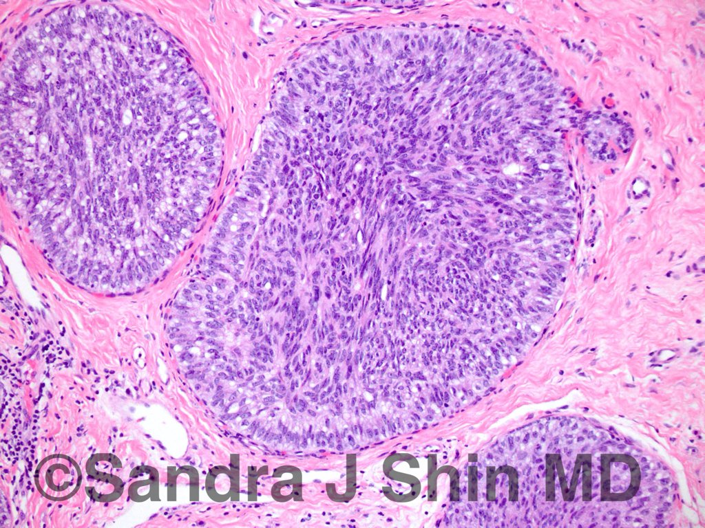
AntiCK 56 has also been found useful in the differential diagnosis of atypical proliferations of the breast The presence of distinct population of CK 56 negative cells supports a diagnosis of ADH, whereas mosaic pattern of CK56 expression favors UDH Preparation for CK5/6 Test Most blood tests do not require any special kind of preparation Cytokeratin 5/6 staining is usually weak or negative for sarcomatoid mesothelioma, the least common and hardesttotreat cell type of the asbestosrelated cancer The marker is also not effective in telling the difference between pleural mesothelioma and squamous cell carcinomasMalignant mesothelioma diagnosis is a panel of two positive and two negative immunohistochemical markers 1 Immunohistochemical staining for Cytokeratin (Pancytokeratin, cat #PDM071) is used for the diagnosis of epithelioid mesothelioma Cytokeratin can be used to highlight stromal invasion of malignant cells 2 Cytokeratin 5/6 (CK 5/6, cat

Triple Negative Breast Cancer Assessing The Role Of Immunohistochemic tt
What is ck 5/6
What is ck 5/6-CK7 / CK in carcinomas of adrenal cortex and prostate ( Mod Pathol 00; ) To distinguish primary lung carcinoma (CK7 / CK) from metastatic colonic carcinoma to lung (CK7 / CK, BMC Cancer 06;631) To help distinguish colon carcinoma (80% are CK) and poorly differentiated prostatic carcinoma (90% are CK) at biopsy Immunohistochemically, the tumor was positive for p63, various types of cytokeratin, CK 5/6, suggesting that the carcinoma had squamous differentiation 911 Negative immunoreactions for TTF1 and PE10 suggest that the tumor had no adenocarcinomatous differentiation 911 The absence of neuroendocrine antigens (chromogranin, synaptophysin


Immunohistochemical And Clinical Characterization Of The Basal Like Subtype Of Invasive Breast Carcinoma Clinical Cancer Research
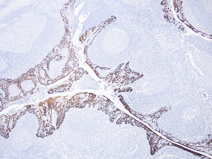
Primary lung adenocarcinomas expressed TTF1 in 90% and napsin A in 84% of the cases, whereas 10% were positive for p63, 7% for CDX2, 2% for CK, and 2% for GATA3 Only 68% of the lung adenocarcinomas were positive for CK7, TTF1, and napsin A and negative for all other markersPRODUCT SPECIFICATIONS Tissue Positive staining tonsil and negative staining liver Fixation Formalin 10%, Phosphate Buffered () Section/Glass Paraffin sections cut at 4 microns on Superfrost™ Plus slides Quality Control Stain Cytokeratin 5/6 (CK5/6) quality control stained slide(s) included Reactivity Guaranteed product specific reactivity for one year from date of By contrast, a negative correlation was observed between CK and CK5/6 In cases with both CK and CK5/6 reactivity, the staining patterns of these two markers were generally reciprocal Although they were not completely mutuallyexclusive, CK tended to be stained on CK5/6negative tumor cells and vice versa
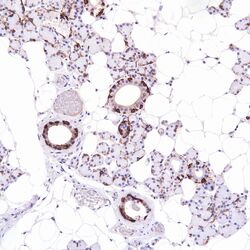
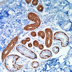
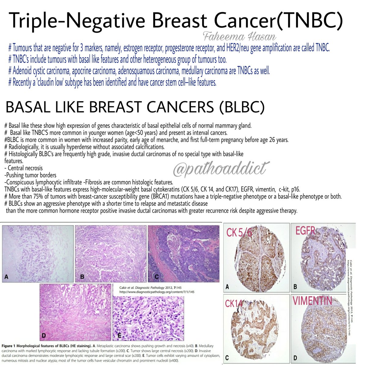
Distribution of p63, cytokeratins 5/6 and cytokeratin 14 in 51 normal and 400 neoplastic human tissue samples using TARP4 multitumor tissue microarray Virchows Arch Int J Pathol 03;–32CK 5/6 expression breast carcinoma implies a 'basal like' molecular phenotype and is associated with poor prognosis This antibody is also used as a component ofCytokeratin 5/6 (CK 5/6) staining basal cells in a benign prostate gland Cytokeratin 5/6 expression in eccrine ducts and coil Note that while both layers of the eccrine duct express CK 5/6, only the basal layer stains positively with CK 5/6 in the eccrine coil Cytokeratin 5/6 expression in
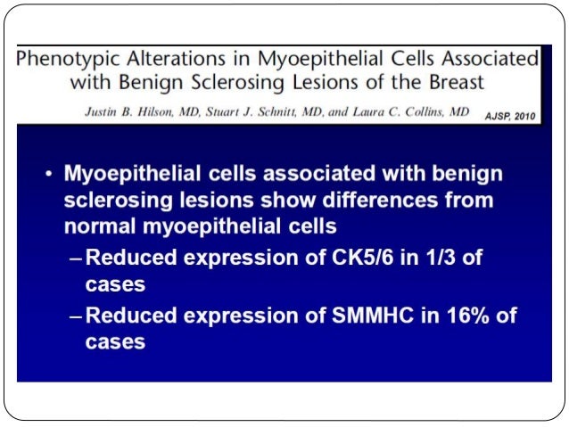
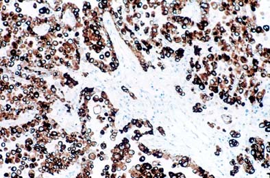
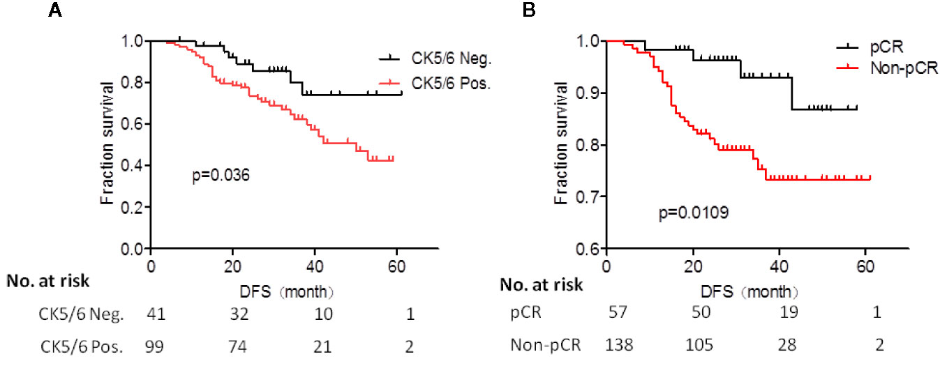
Cytokeratin 5/6 (CK5/6) is an intermediate filament of the group of highmolecularweight cytokeratins and is consistently expressed in stratified epithelia and derived neoplasm such as squamous carcinoma Furthermore, CK5/6 is expressed in the basalmyoepithelial layer of prostate, breast, and salivary glands ,An association with ERα expression could not be detected but the combination of cytokeratin 5/6 IRS = 12 and ERα negativity resulted in a significant negative prognostic marker (overall survival P = 003 and progressionfree survival P < 0001) We substantiate cytokeratin 5/6 protein expression as a frequent feature of highgrade serousFor CK5 staining 96% of ADH/DCIS cases were negative or focally positive, whereas all 32 UDH cases had diffuse staining The combination of ER and CK5 increased the sensitivity (94% to 97%) For PR staining 11 of 23 ADH cases (48%), 6 of 10 DCIS cases (60%), and 4 of 32 UDH cases (13%) showed diffuse staining l2 staining showed no


1


Prognostic Impact Of Egfr And Cytokeratin 5 6 Immunohistochemical Expression In Triple Negative Breast Cancer Sciencedirect
Breast cancer cells taken out during a biopsy or surgery will be tested to see if they have certain proteins that are estrogen or progesterone receptors When the hormones estrogen and progesterone attach to these receptors, they fuel the cancer growth Cancers are called hormone receptorpositive or hormone receptornegative based on whether or not they have these All cases were Ecadherin negative CK5/6 was positive in 14 (17%) of cases and was entirely negative in the remaining 68 cases (%) In 8 of the 14 CK5/6 cases, staining was diffuse and intense In the remaining 6 cases, staining was patchy (>1 lowpower field between positive areas) but still of high intensity p63 and cytokeratin (CK) 5/6 are both considered to be markers of basal and squamous cell differentiation in several normal epithelia and human tumors 1, 2 Previous studies have indicated that p63 is important for epithelial proliferation and differentiation, 2 and studies using knockout mice have suggested that p63 plays a key role in maintaining progenitor cell
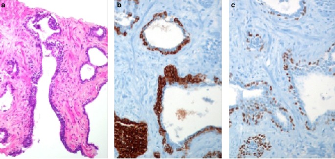


Diagnostic Utility Of A P63 A Methyl Coa Racemase P504s Cocktail In Atypical Foci In The Prostate Modern Pathology


Ck 5 6 Expression In Breast Ductal Carcinomas A Triple Negative Download Scientific Diagram
Of the lungis generally negative6 transitional cell carcinoma5 mesothelium and mesotheliomais frequently Initially K5 was suggested as a marker for mesotheliomaConclusion CK 5/6 expression breast carcinoma implies a 'basal like' molecular phenotype and is associated with poor prognosis This antibody is also used as a component of panels to differentiate benign and malignant breast lesionsAll of the 10 ACs were negative for p63 and most of them (8/10) were negative for CK5/6 p63 and CK 5/6 seem to be useful for differentiating AC and SCLC from SCC with 100% specificity and % sensitivity, % specificity and 79% sensitivity, respectively


Immunohistochemistry In Prostate Pathology Pdf Free Download
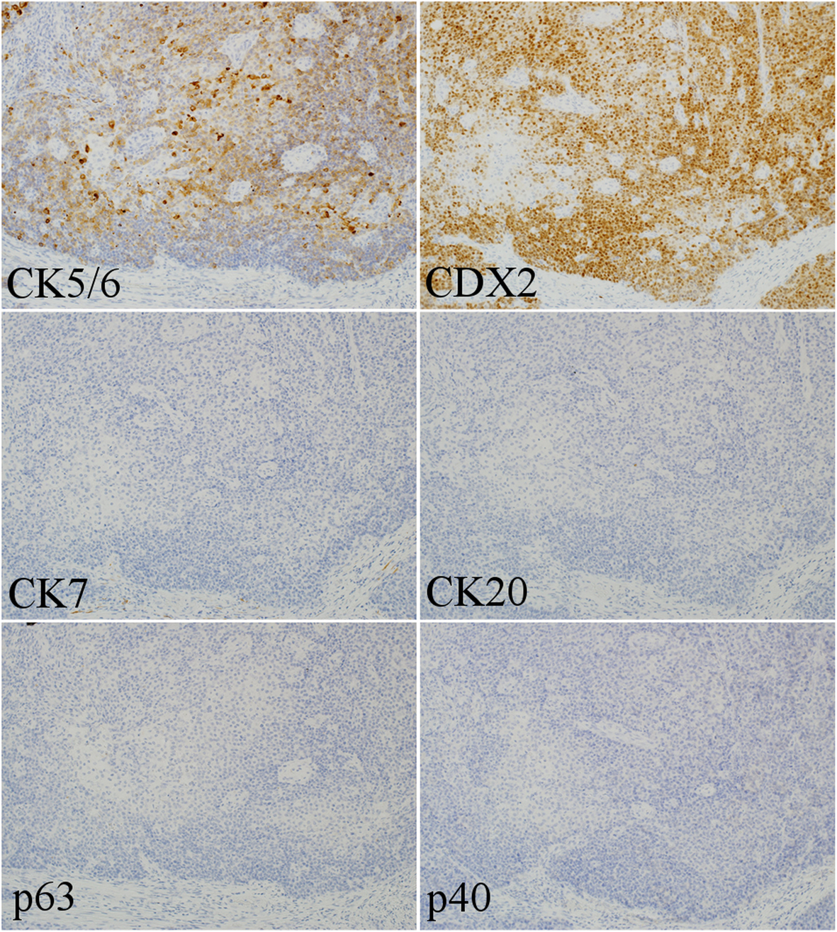


The Origin Of P40 Negative And Cdx2 Positive Primary Squamous Cell Carcinoma Of The Stomach Case Report World Journal Of Surgical Oncology Full Text
Most p63expressing tumors diffusely and strongly express CK18 at levels similar to benign luminal cells (case 1 and 3), although some show weaker diffuse expression (case 2) Similarly, most are entirely negative for CK5/6 (cases 2 and 3), although a minority weakly and focally express CK5/6 (case 1) All images are at 0 × magnification For CK5/6, 94% of FEA, ADH, DCIS, and LCIS revealed negative stainings, but all of the UDH demonstrated diffuse stainings For CK903, 90% of FEA, ADH, and DCIS were negative to focally positive, and all of the UDH and 90% of LCIS were diffusely positive Considered a marker of or associated with the basal phenotype (also CK 5/6, CK 14) of invasive or in situ ductal carcinoma of breast (Mod Pathol 06;) Sensitive marker of sentinel nodal metastases by RTPCR in oral squamous


Pdf Prognostic Impact Of Egfr And Ck 5 6 As Basal Markers In Triple Negative Breast Cancers Semantic Scholar
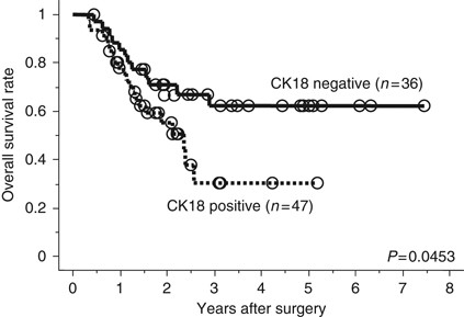


Cytokeratins 18 And 8 Are Poor Prognostic Markers In Patients With Squamous Cell Carcinoma Of The Oesophagus British Journal Of Cancer
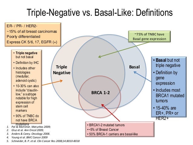
In the ER negative cases, 60% and 45% were positive for CK5/6 and CK14, respectively, whereas 393% and 303% of PR negative cases showed positivity for CK5/6 and CK14, respectively In contrast, only 303% of ER positive tumors were positive for CK5/6 and CK14 expression None of the cases which were PR positive showed expression of basal CKs IHC staining for CK 5/6, CK and CD 44 was analysed in 211 patients with non‐muscle‐invasive papillary UTUC Staining was classified as showing a negativeHigher grade papillary urothelial carcinoma, on the other hand, reveals CK expression in the entire thickness of the urothelium with the absence of
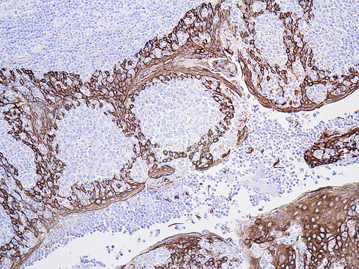


Cytokeratin 5 6 Ck5 6 Histology Control Slides
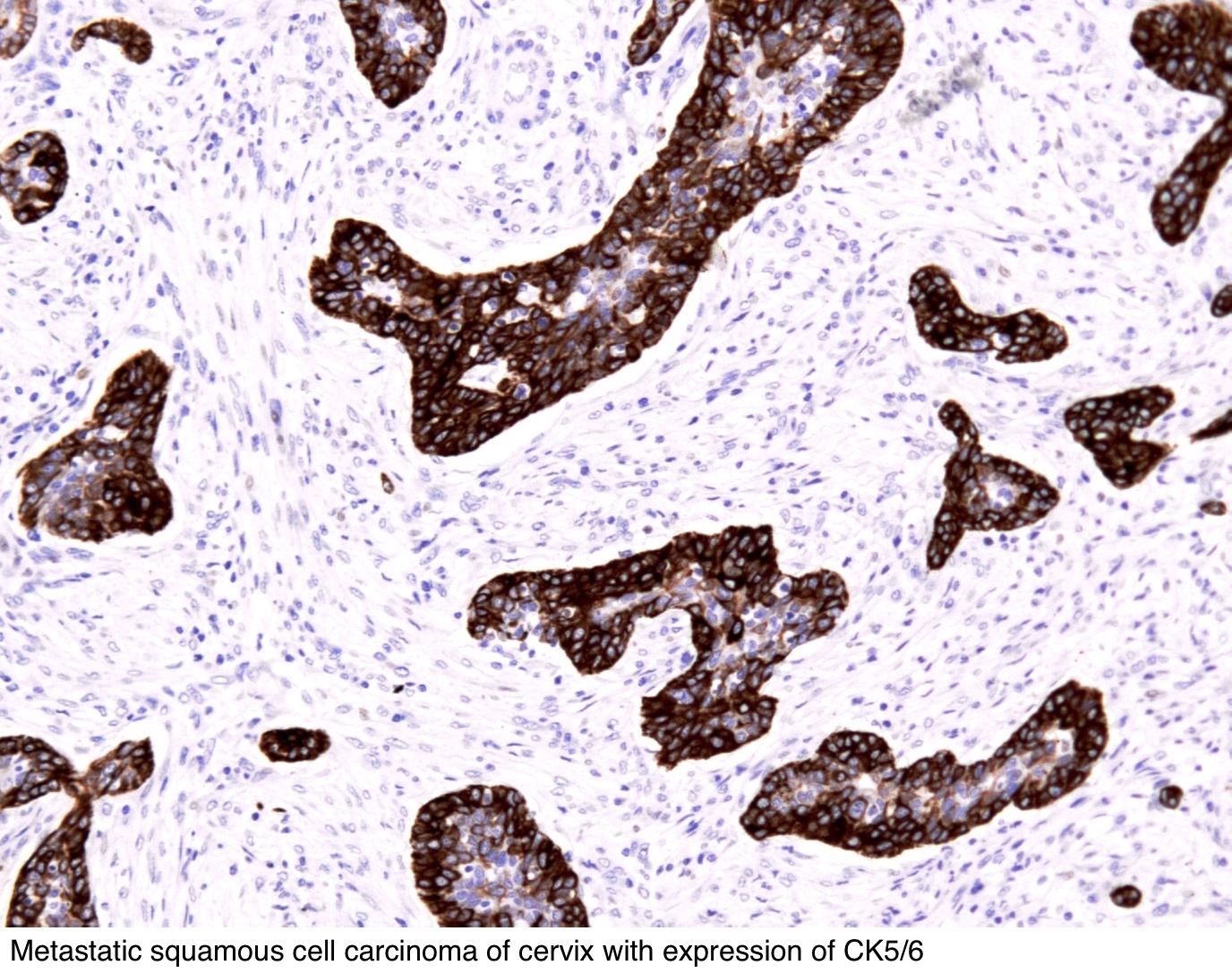


Pathology Outlines Cytokeratin 5 6 And Ck5
TTF1, CK 5/6 and p63 seem to be useful for differentiating adenocarcinoma from squamous cell carcinoma with 100% specificity, 100% sensitivity and 100% specificity, 97% sensitivity and 87% Kelly, late s, early 30s, blue eyes, short, silly dancer, so much fun!!Product Description Studies have shown Cytokeratin 5/6 antibody reacts with human epidermis and nonkeratinizing epithelium It has also been shown to react with cytokeratin 6, weakly with cytokeratin 4, and does not react with cytokeratins 1, 7, 8, 10, 13, 14, 18 and 19 CK5/6 has been shown to express in the vast majority of squamous cell


1


Pdf Value Of P63 And Cytokeratin 5 6 As Immunohistochemical Markers For The Differential Diagnosis Of Poorly Differentiated And Undifferentiated Carcinomas
Aims Immunohistochemical (IHC) staining for cytokeratin (CK) 5/6, Staining was classified as showing a negative, positive or normal pattern We found that CK5/6negative, CD44negative and CKpositive tumours were distinctly highrisk subgroups that were associated with high grade (CK5/6negative, P < 0001;Coexpression of p63 and CK5/6 had a sensitivity of 077 and a specificity of 096 for squamous cell carcinomas Increasing the minimal criterion of positive immunostaining for both markers to more than 50% of immunoreactive tumor cells resulted in a specificity of 099, although the sensitivity diminished to 0661 Downey P, Cummins R, Moran M, et al If it's not CK5/6 positive, TTF1 negative it's not a squamous cell carcinoma of lung Apmis –529, 08 2 Kargi A, Gurel D, Tuna B The diagnostic value of TTF1, CK 5/6, and p63 immunostaining in classification of lung carcinomas Appl Immunohistochem Mol Morphol –4, 07 3
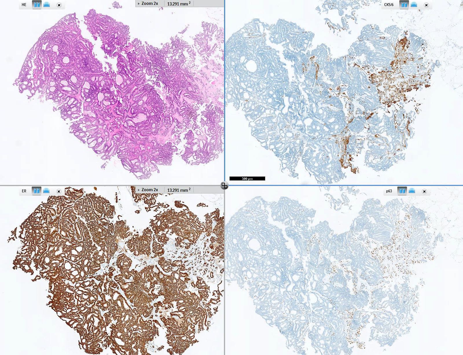


Pathology Outlines Cytokeratin 5 6 And Ck5
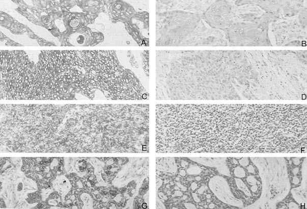


Expression Of Cytokeratin 5 6 In Epithelial Neoplasms An Immunohistochemical Study Of 509 Cases Modern Pathology
Use Prostatic adenocarcinoma versus highgrade prostatic intraepithelial neoplasia (HGPIN) CK5/6 marks the basal cells present in HGPIN, absent in cancer Urothelial cell carcinoma versus prostatic adenocarcinoma Positive in UCC, negative in prostate adenocarcinoma Abstract Breast Cancer Res Treat (09) –663 DOI /sy LETTE R T O T HE EDI T OR The immunohistochemically ''ERnegative, PRnegative, HER2negative, CK5/6negative, and HER1negative'' subgroup is not a surrogate for the normallike subtype in breast cancer KeDa Yu Æ ZhenZhou Shen Æ ZhiMing Shao Received 17All of the 10 ACs were negative for p63 and most of them (8/10) were negative for CK5/6 p63 and CK 5/6 seem to be useful for differentiating AC and SCLC from SCC with 100% specificity and % sensitivity, % specificity and 79% sensitivity, respectively
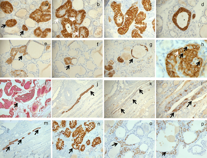


Thyroid Solid Cell Nests Usefulness Of Cytokeratin 5 6 Springerlink
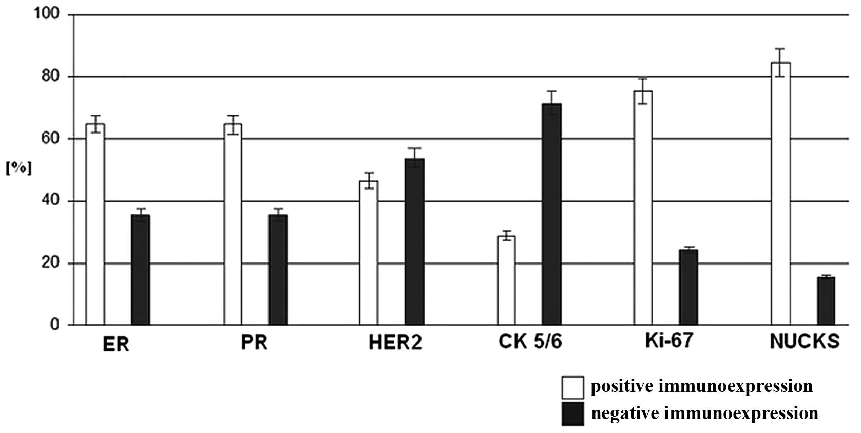


Immunohistochemical Study Of Nuclear Ubiquitous Casein And Cyclin Dependent Kinase Substrate 1 In Invasive Breast Carcinoma Of No Special Type
By Rodney T Miller, MD Cytokeratin 5/6 (CK 5/6) is a very useful antibody in diagnostic Surgical Pathology One of the most important aspects of this marker is that it characteristically stains squamous carcinomas strongly and diffusely, but generally stains adenocarcinomas focally, weakly, or not at all As such, it can be used as a marker of squamous differentiationThere is often focal and patchy basal cells staining with occasional glands being totally negative for basal cells (Figures 34) (7) CK HMW, CK 5/6 and p63 A pitfall in the use of immunohistochemistry for the di agnosis of prostate adenocarcinoma is false positive staining for basal cell markers This can occur in several patterns As brought out in our results none of the SNUC was positive for either CK 5/6 orp40 In comparison, the undifferentiated nasopharyngeal carcinoma and poorly differentiated squamous cell carcinoma both exhibited for CK 5/6 and p40 in 90% and 100% of cases, respectively None of the ONB was positive for either of studied markers
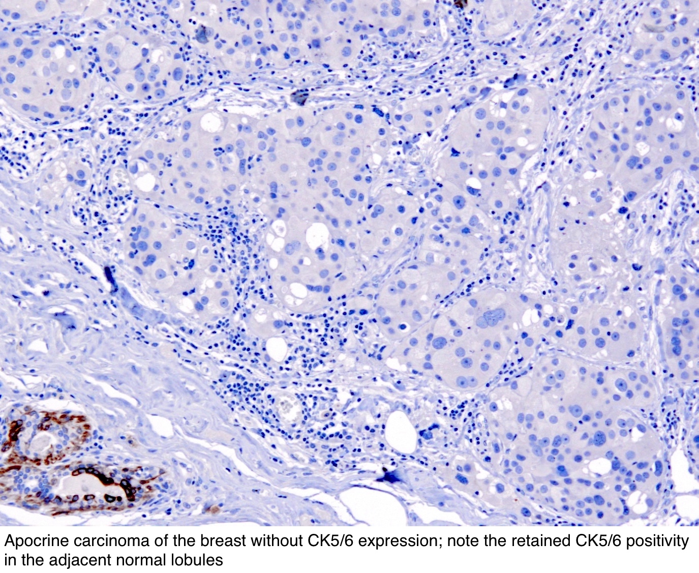


Pathology Outlines Cytokeratin 5 6 And Ck5
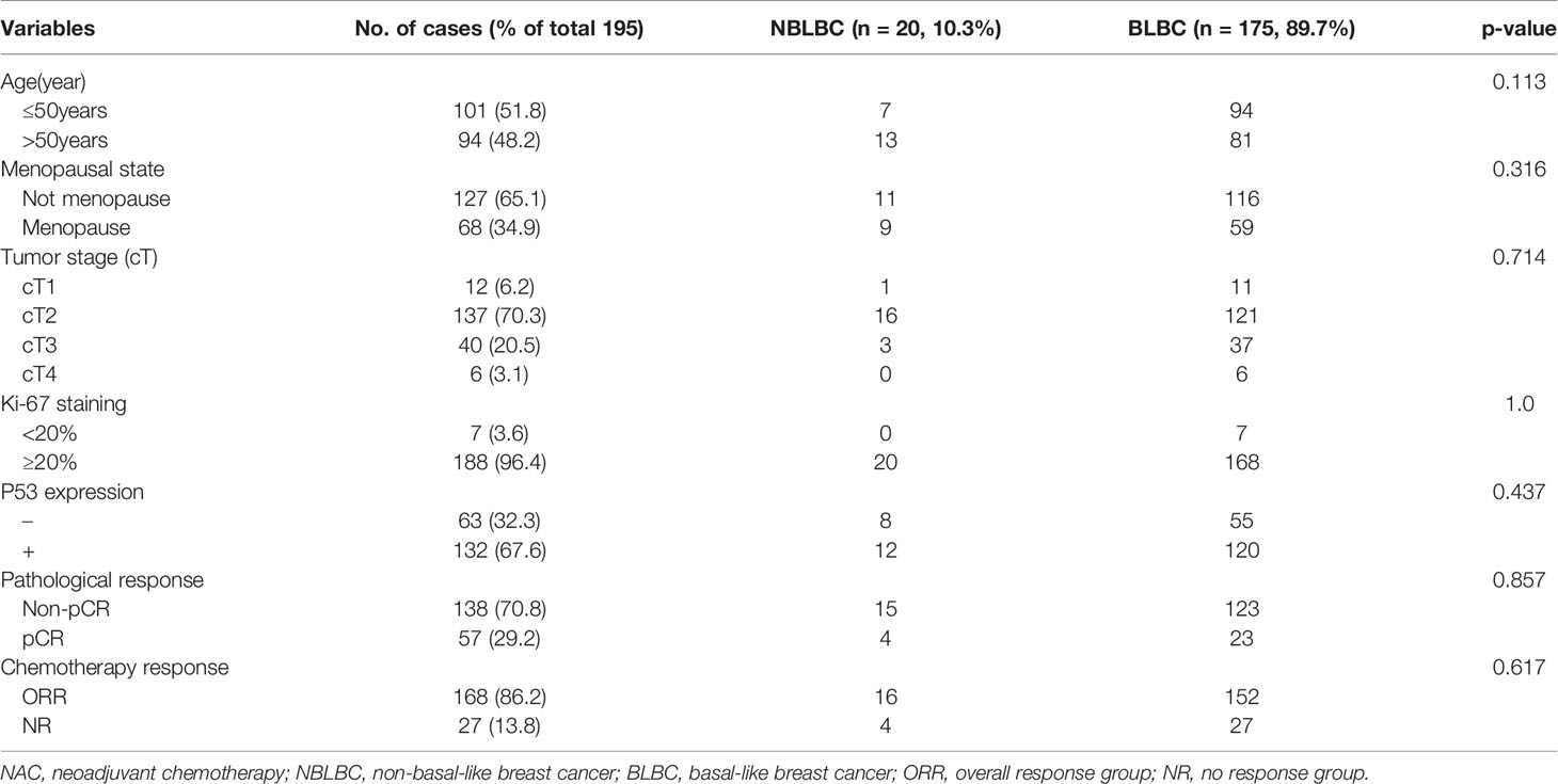


Frontiers Analysis Of Ck5 6 And Egfr And Its Effect On Prognosis Of Triple Negative Breast Cancer Oncology
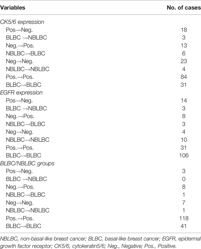
1 Downey P, Cummins R, Moran M, et al If it's not CK5/6 positive, TTF1 negative it's not a squamous cell carcinoma of lung Apmis –529, 08 2 Kargi A, Gurel D, Tuna B The diagnostic value of TTF1, CK 5/6, and p63 immunostaining in classification of lung carcinomas Appl Immunohistochem Mol Morphol –4, 07 3 Positive CK5/6 expression was noted in 8% (12 cases) of TNBC while 24% (4 cases) showed focal positive ( Common antibodies are directed against both cytokeratin 5 and 6 Major partner of CK5 is CK14, while CK6 pairs with CK16/17 (Exp Cell Res 07;, Eur J Cell Biol 04;735) CK5 also detected by antibodies against high molecular weight cytokeratins (such as 34βE12, also known as K903) and panCK markers (such as CK AE1/A)
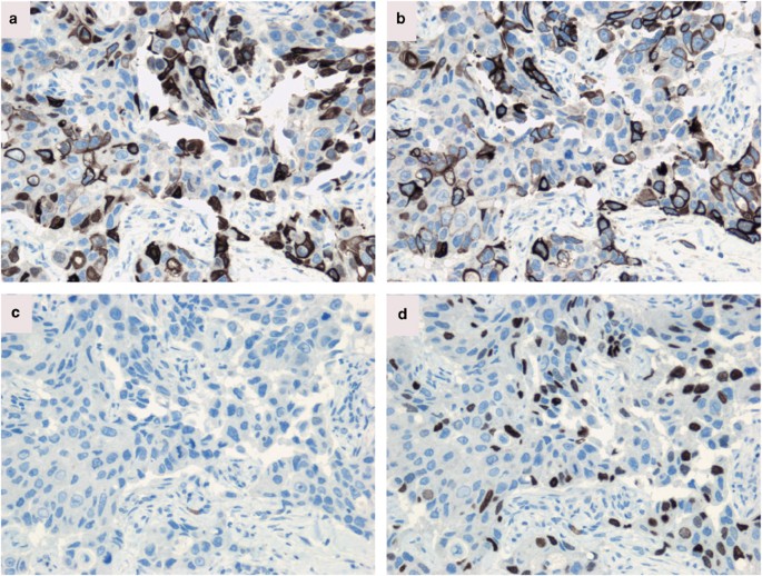


Cytokeratin 5 14 Positive Breast Cancer True Basal Phenotype Confined To Brca1 Tumors Modern Pathology



P53 Expression In Triple Negative Breast Carcinomas Evidence Of Age Related And Racial Differences
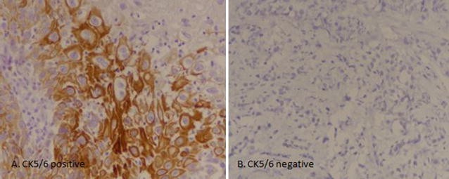
Cyclin D1 and CK5/6 staining could be used in concert to distinguish between the diagnosis of papilloma (Cyclin D1 3700%, CK 5/6 negative) In the lung, distinguishing epithelioid mesothelioma (CK5/6 positive in %) from lung adenocarcinoma (CK5/6 negative in 85%) Positive CK5/6 expression was noted in 63% (8 cases) and 134% (17 cases) revealed focal positive CK 5/6 expression On the other hand, 803% (102 cases) showed negative CK5/6 staining Significant association of CK5/6 expression was noted with tumor grade and muscularis propria invasion, however no significant association was noted with Cytokeratin 5/6 has been shown to be present in the luminal epithelial proliferating cells of usual ductal hyperplasia in % to 100% of cases 12,13,15 Conversely, the luminal cells of ADH and lowgrade DCIS tend to be negative or show only scattered positivity 12–15 Our results also support these observations


Cytokeratin 5 6 Ck 5 6 Pathology Resident Wiki Fandom


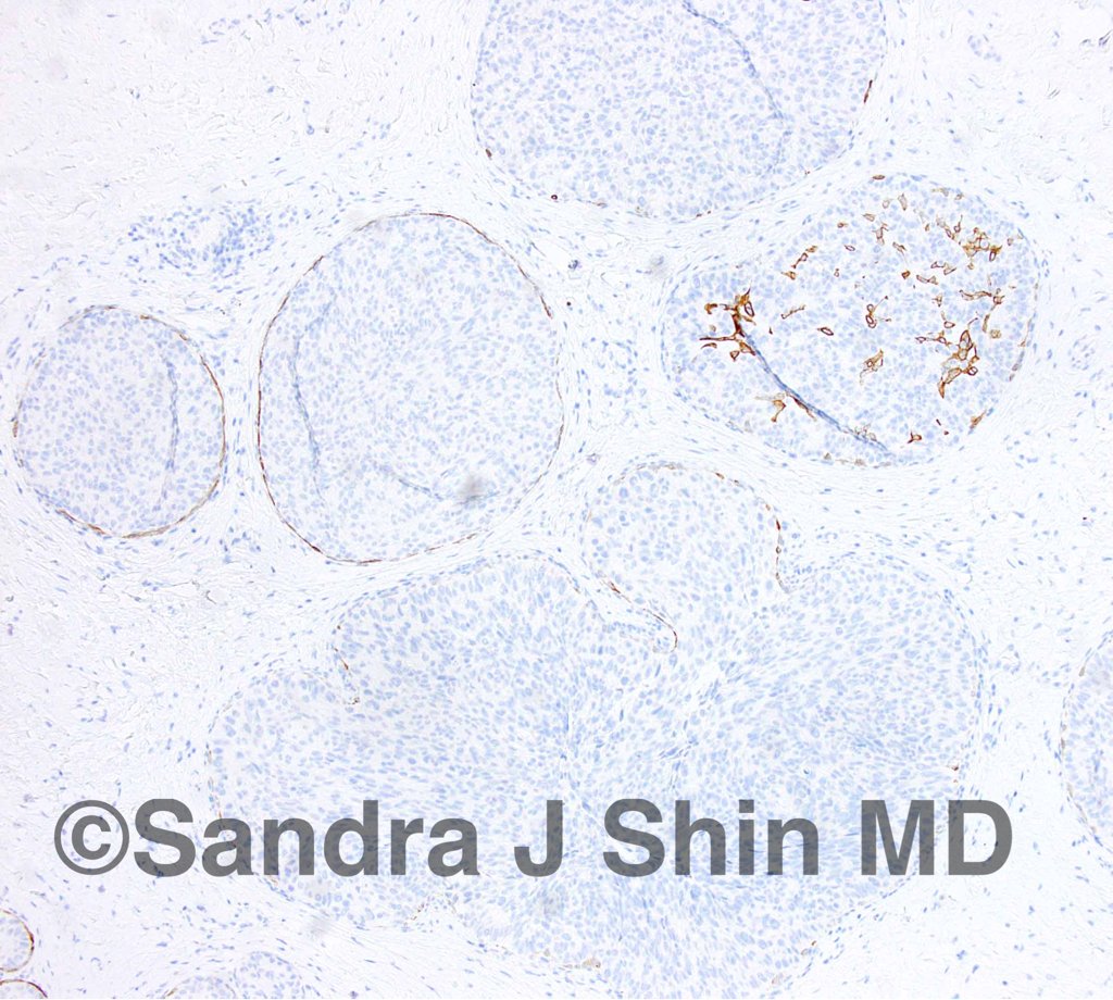
Sandra J Shin Md Mba Dcis With Spindle Cell Morphology Negative Ck 5 6 Helps To Distinguish From Florid Usual Ductal Hyperplasia T Co Vfclbmz3o3
Squamous cell carcinomas and adenocarcinomas are the most common types of cervical cancer Compared to squamous cell carcinomas, adenocarcinomas are more common in younger women and have a poorer prognosis Yet, so far, no useful biomarkers have been developed for these two types of cancer In the following study, we examined the combination of cytokeratin 5/6We hung out Saturday second set and briefly last night and I wiffed on asking for your number Your friend is a tall brunette named Lauren who was also rad as hell Page side roughly near that damn light 🤙 You're super cute and I had a lot of fun, my friends all Our results show that Cyclin D1 and CK 5/6 staining could be used in concert to distinguish between the diagnosis of papilloma (Cyclin D1 < 4%, CK 5/6 positive) or papillary carcinoma (Cyclin D1 > 3700%, CK 5/6 negative)


Ros1 Expression And Translocations In Non Small Cell Lung Cancer Clinicopathological Analysis Of 1478 Cases Warth 14 Histopathology Wiley Online Library


Ihc In Breast Pathology


1


Unclineberger Org Peroulab Wp Content Uploads Sites 1008 19 06 Aug 15 Basal Like By Ihc Nielsen Ccr 04 Pdf


Cytokeratin 5 6 Ck 5 6 Pathology Resident Wiki Fandom


Scielo Brasil Secretory Carcinoma Of The Breast Commonly Exhibits The Features Of Low Grade Triple Negative Breast Carcinoma A Case Report With Updated Review Of Literature Secretory Carcinoma Of The


2


Prognostic Impact Of Egfr And Cytokeratin 5 6 Immunohistochemical Expression In Triple Negative Breast Cancer Sciencedirect
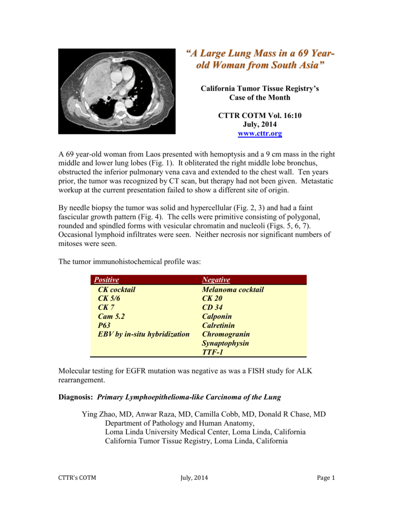


Cotm0714 Final California Tumor Tissue Registry
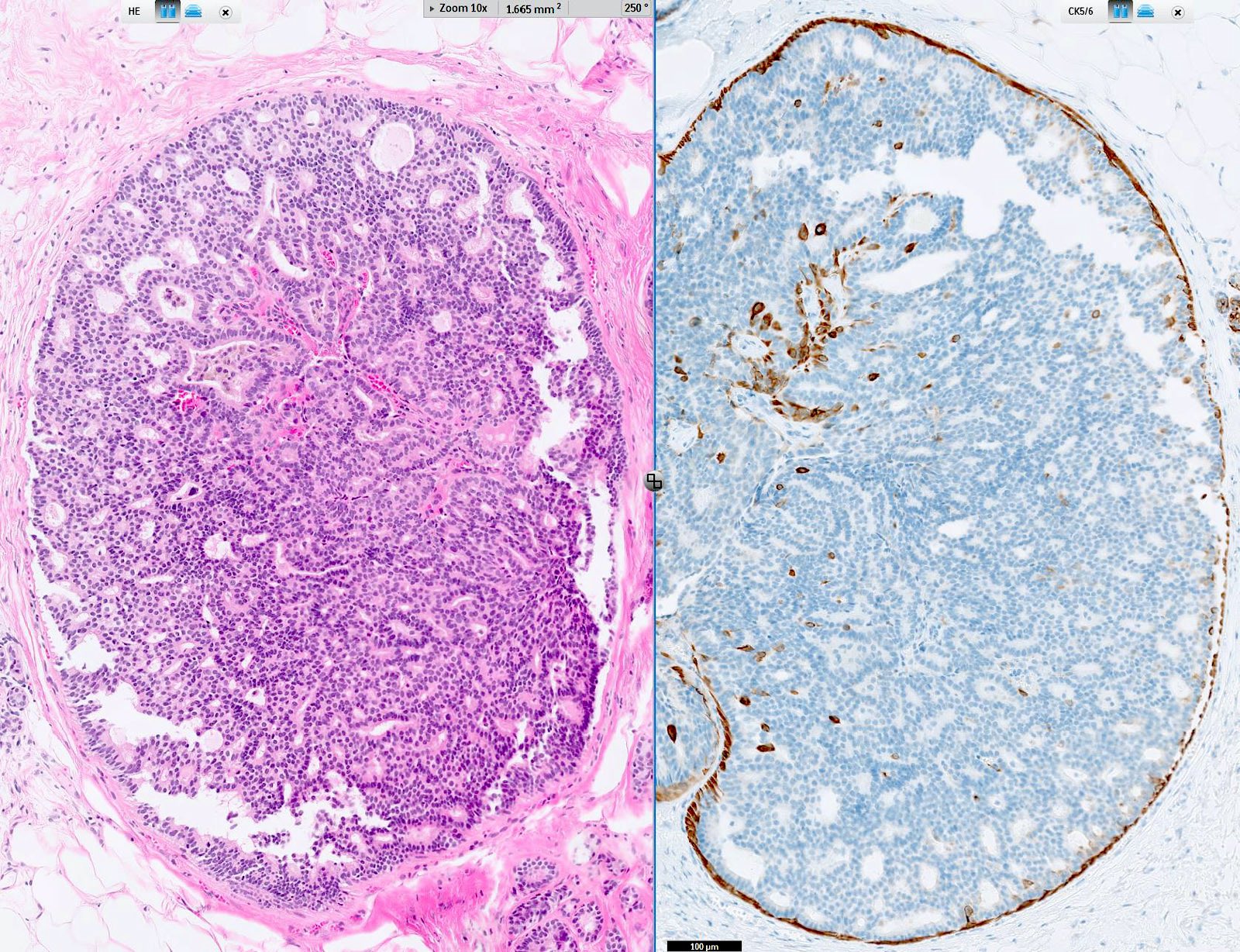


Pathology Outlines Cytokeratin 5 6 And Ck5


Ck 5 6 Expression In Breast Ductal Carcinomas A Triple Negative Download Scientific Diagram
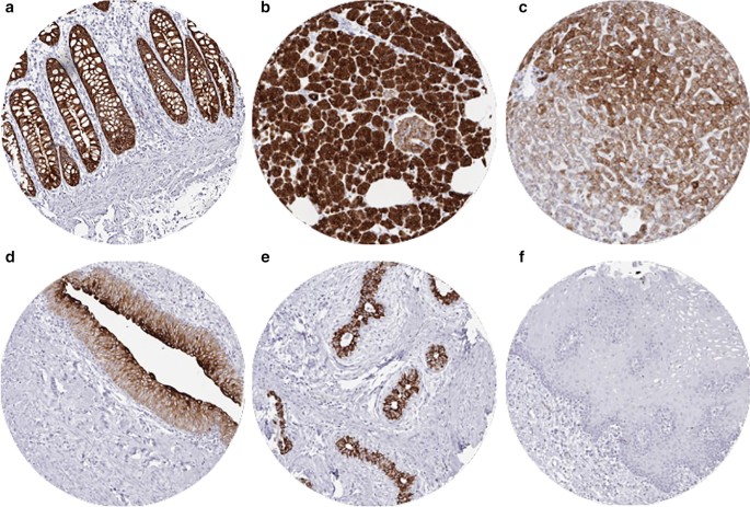


Diagnostic And Prognostic Impact Of Cytokeratin 18 Expression In Human Tumors A Tissue Microarray Study On 11 952 Tumors Molecular Medicine Full Text


Pdf Basal Cytokeratin As A Potential Marker Of Low Risk Of Invasion In Ductal Carcinoma In Situ Semantic Scholar


Triple Negative Breast Cancer At The Jos University Teaching Hospital Mandong Bm Emmanuel I Vandi Kb Yakubu D Ann Trop Pathol


Www Ajol Info Index Php Mjz Article View


Hematoxylin Eosin He Staining And Immunohistochemistry The Tumor Download Scientific Diagram
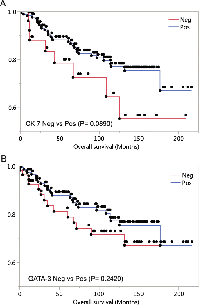


Cytokeratin 7 Negative And Gata Binding Protein 3 Negative Breast Cancers Clinicopathological Features And Prognostic Significance Bmc Cancer Full Text
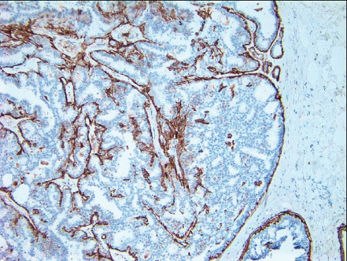


Papillary Lesions Oncohema Key
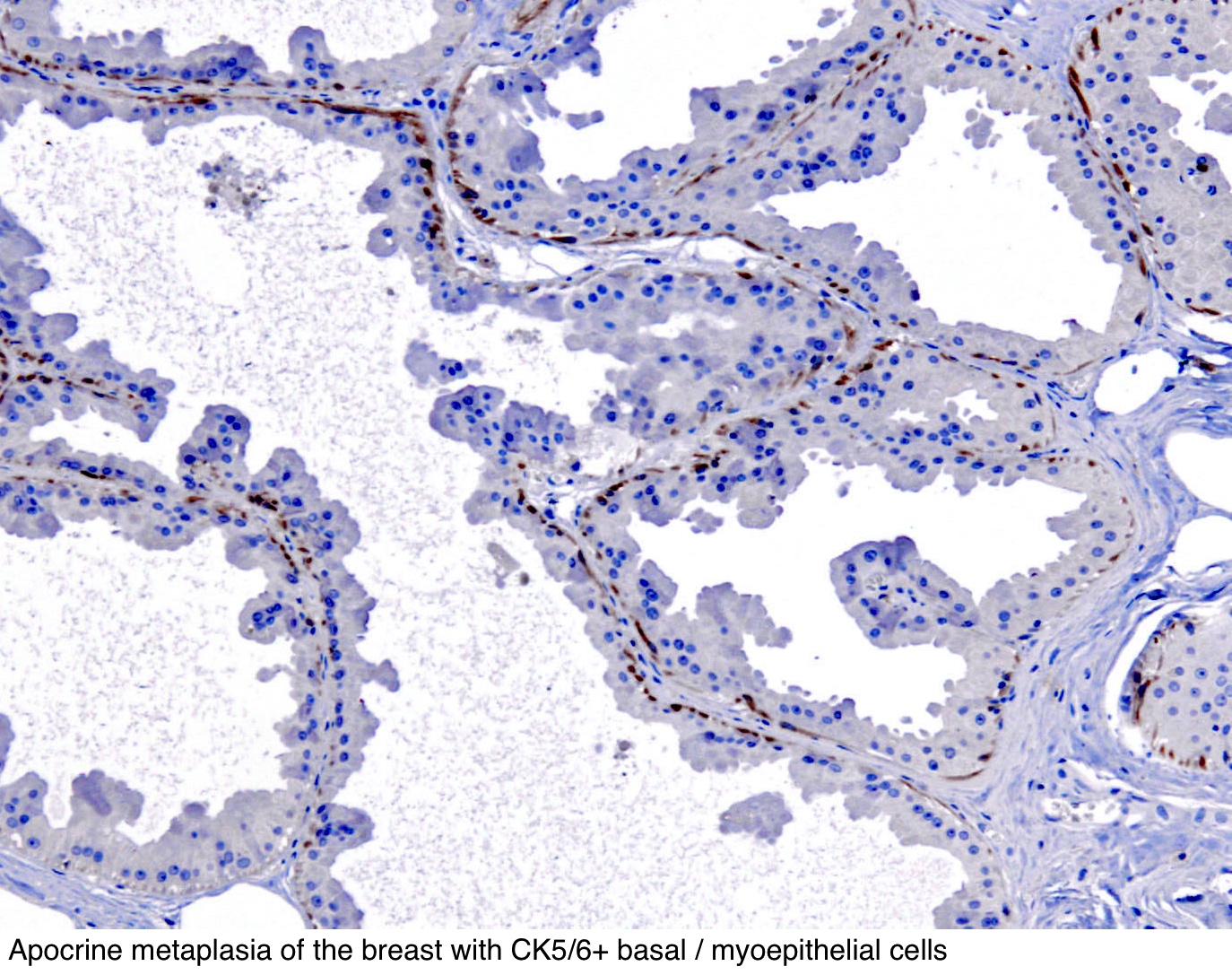


Pathology Outlines Cytokeratin 5 6 And Ck5


Triple Negative Breast Cancer Assessing The Role Of Immunohistochemic tt
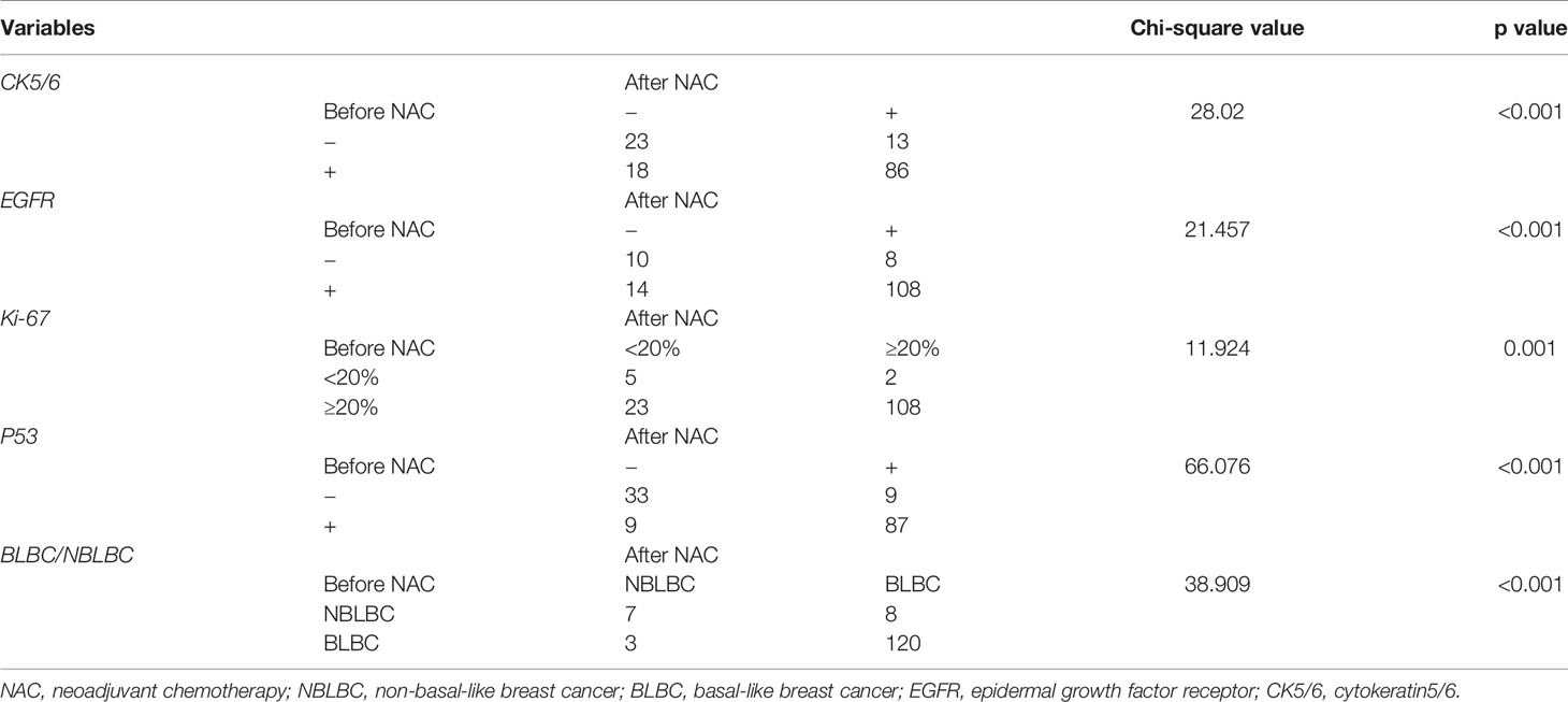


Frontiers Analysis Of Ck5 6 And Egfr And Its Effect On Prognosis Of Triple Negative Breast Cancer Oncology
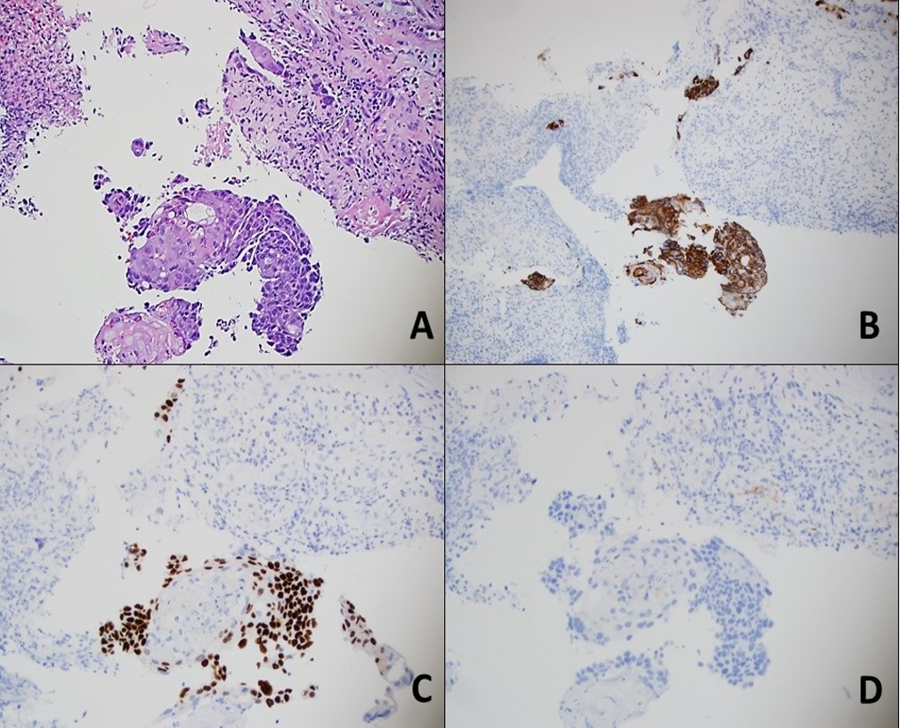


Cureus Primary Gastric Squamous Cell Carcinoma


2


Malignant Mesothelioma A Histomorphological And Immunohistochemical Study Of 24 Cases From A Tertiary Care Hospital In Southern India


Biological Significance Of Gata3 Cytokeratin Cytokeratin 5 6 And P53 Expression In Muscle Invasive Bladder Cancer


Pathology Outlines Cytokeratin 5 6 And Ck5


Jaypeedigital Ebook Reader


Cytokeratin 5 6 Expression In Bladder Cancer Association With Clinicopathologic Parameters And Prognosis Bmc Research Notes Full Text


Faheema Hasan Md Characteristics Of Triple Negative Breast Cancers Pathology Breastpath Pathboards Pembeoltulu Kriyer68 Suraksharaob Binxu16 Appy Vashi Geronimojrlapac Smlungpathguy Vijaypatho T Co Pxbvs754se


Frontiers Analysis Of Ck5 6 And Egfr And Its Effect On Prognosis Of Triple Negative Breast Cancer Oncology


Frontiers Analysis Of Ck5 6 And Egfr And Its Effect On Prognosis Of Triple Negative Breast Cancer Oncology


Cytokeratin 5 6 Ck5 6 Histology Control Slides


Pathology Outlines Cytokeratin 5 6 And Ck5


Immunohistochemical And Clinical Characterization Of The Basal Like Subtype Of Invasive Breast Carcinoma Clinical Cancer Research


Indian Journal Of Dermatology Venereology And Leprology Practical Immunohistochemistry Of Epithelial Skin Tumor


Triple Negative Breast Cancer
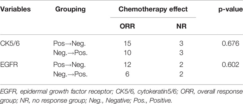


Frontiers Analysis Of Ck5 6 And Egfr And Its Effect On Prognosis Of Triple Negative Breast Cancer Oncology
-Flow-Cytometry-NBP2-52444-img0001.jpg)


Cytokeratin 5 Antibody 3d6d4 Nbp2 Novus Biologicals


Prognostic Impact Of Egfr And Cytokeratin 5 6 Immunohistochemical Expression In Triple Negative Breast Cancer Sciencedirect


Sandra J Shin Md Mba Dcis With Spindle Cell Morphology Negative Ck 5 6 Helps To Distinguish From Florid Usual Ductal Hyperplasia T Co Vfclbmz3o3


Highlights In The Management Of Breast Cancer How


Epidemiology Definition Biology Basal Like The Triple Negative Breast Cancer Estrogen Receptor Er Negative Progesterone Receptor Pr Negative Ppt Download
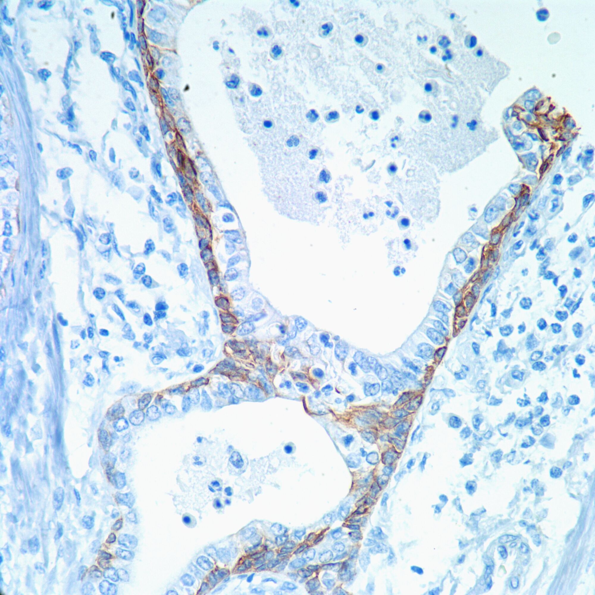


Cytokeratin 5 6 Ck 5 6 Pathology Resident Wiki Fandom


Expression Of P63 And Ck5 6 In Early Stage Lung Squamous Cell Carcinoma Is Not Only An Early Diagnostic Indicator But Also Correlates With A Good Prognosis Ma 15 Thoracic Cancer


Triple Negative Breast Cancer Risk Factors To Potential Targets Clinical Cancer Research


Immunohistochemical Differential Diagnoses In Urological Pathology An Overview


Highlights In The Management Of Breast Cancer How


Pdf Prognostic Impact Of Egfr And Ck 5 6 As Basal Markers In Triple Negative Breast Cancers Semantic Scholar


Academic Oup Com Ajcp Article Pdf 130 5 724 Ajcpath130 0724 Pdf


Her 2 Neu Positive Ihc Stain Slide For Ck 5 6 Receptor Showing Download Scientific Diagram


Figure 2 From Cinical Significance Of Androgen Receptor Ck 5 6 Ki 67 And Molecular Subtypes In Breast Cancer Semantic Scholar


2


Cytokeratin 5 6 Ck5 6 Histology Control Slides
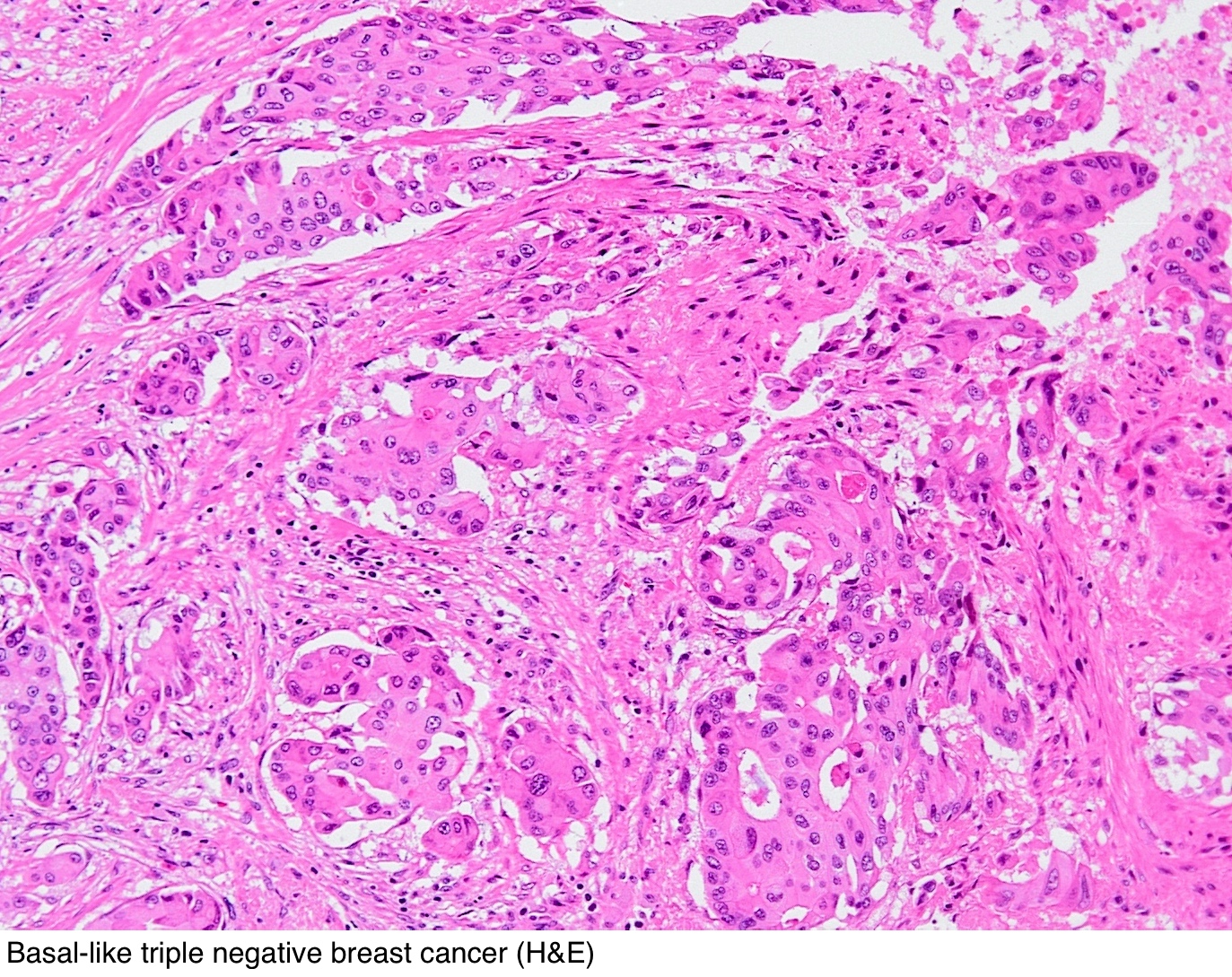


Pathology Outlines Cytokeratin 5 6 And Ck5
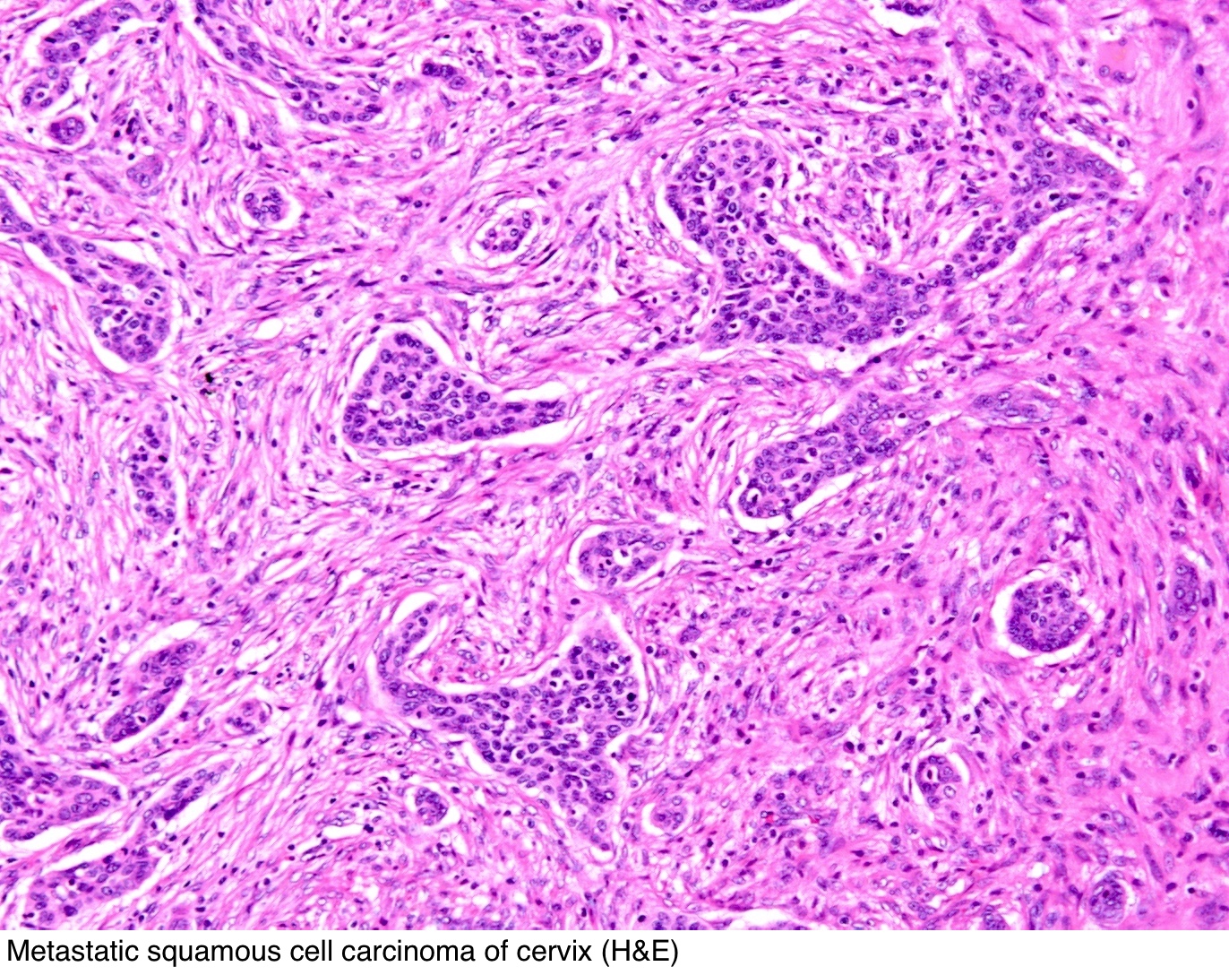


Pathology Outlines Cytokeratin 5 6 And Ck5
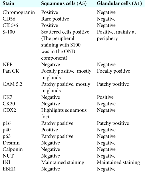


Mixed Olfactory Neuroblastoma And Neuroendocrine Carcinoma An Unusual Case Report And Literature Review Surgical Neurology International


Figure 3 From Another Review On Triple Negative Breast Cancer Are We On The Right Way Towards The Exit From The Labyrinth Semantic Scholar
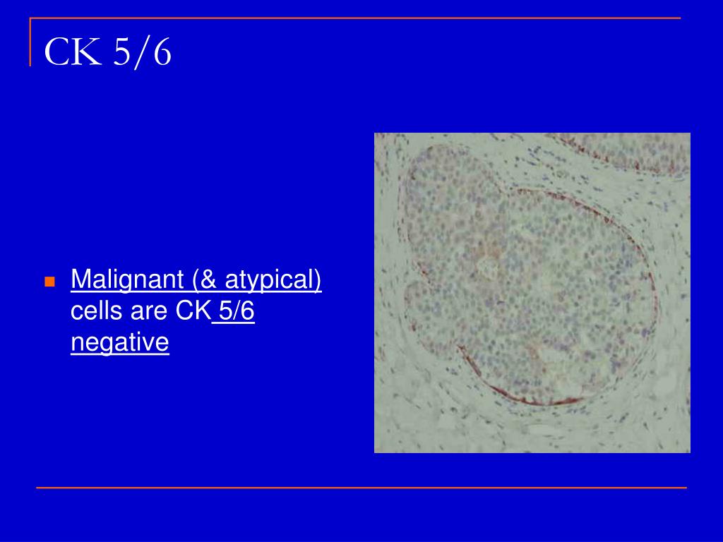


Ppt New Issues In The Interpretation Of Breast Biopsies Powerpoint Presentation Id


Q Tbn And9gctuaeubeofickfpn5 Lfmiu2rtllphz Iclkv2zftgxliqt Avr Usqp Cau


Molecular Classification Of Triple Negative Breast Cancer 103 110 Download Scientific Diagram
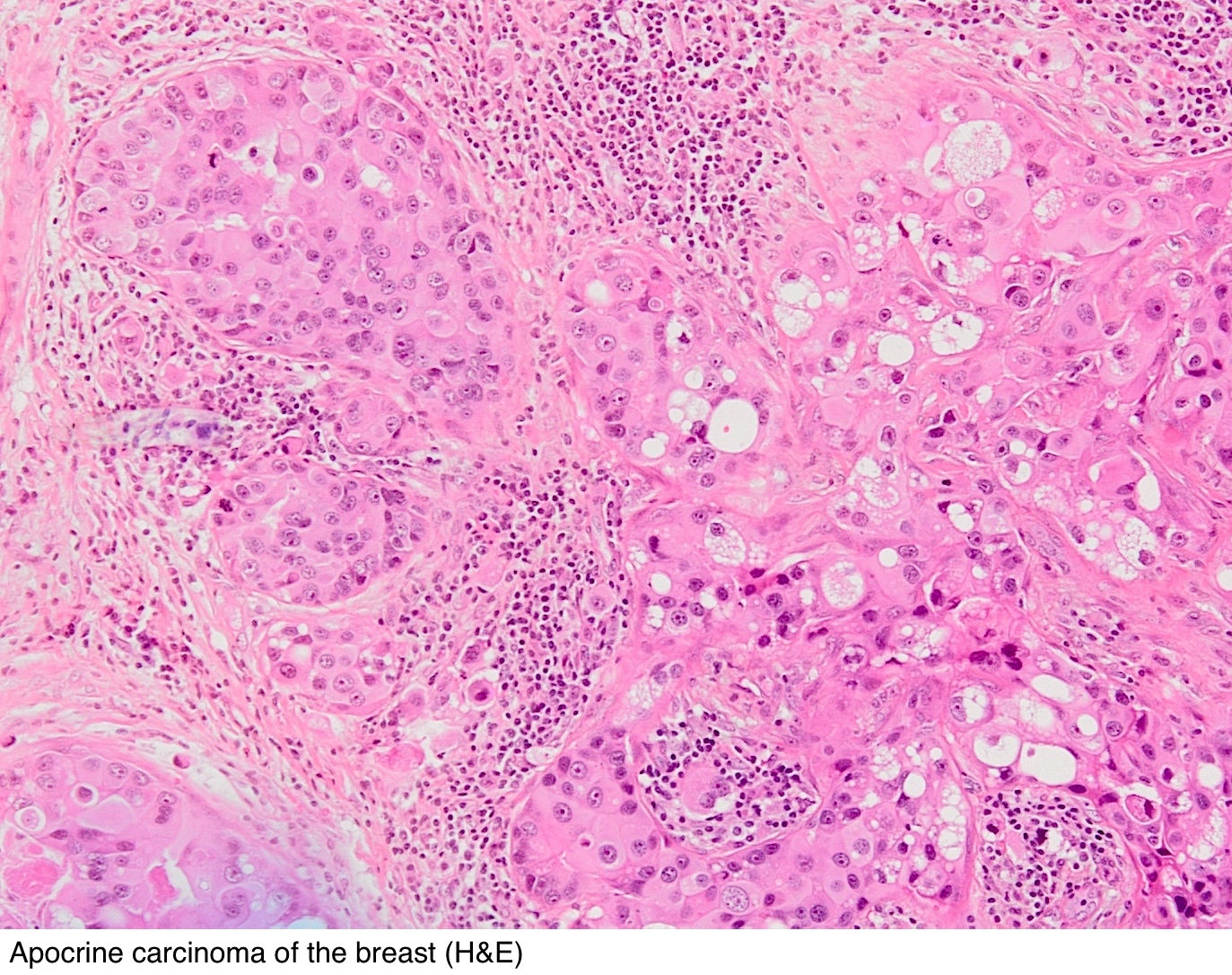


Pathology Outlines Cytokeratin 5 6 And Ck5


Prognostic Impact Of Egfr And Cytokeratin 5 6 Immunohistochemical Expression In Triple Negative Breast Cancer Sciencedirect
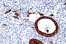


Cytokeratin Wikipedia


Lung Squamous Cell Carcinoma Associated With Hypoparathyroidism With S Ott
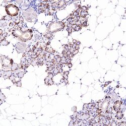


Cytokeratin 5 6 Ck 5 6 Pathology Resident Wiki Fandom
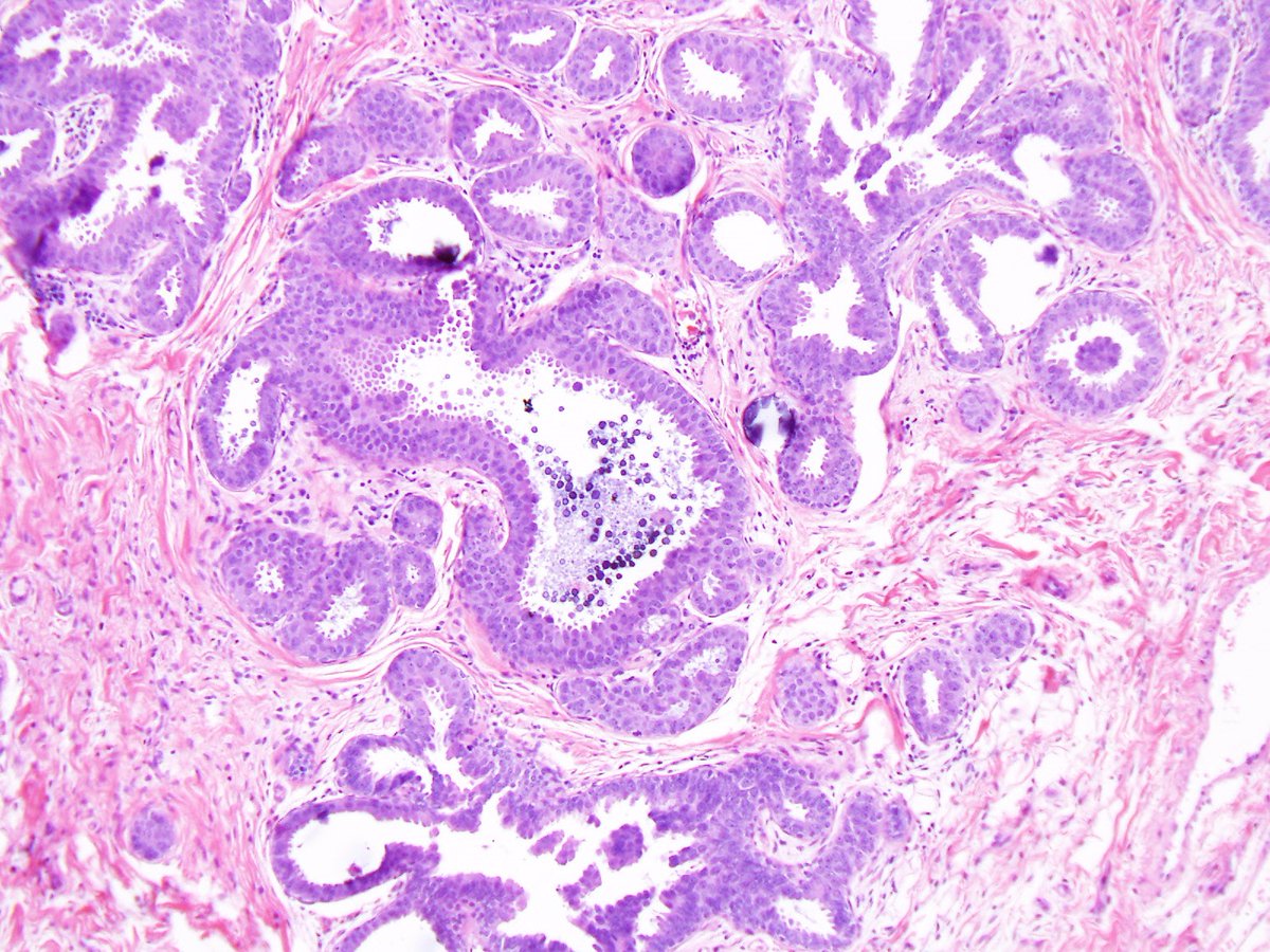


Reza Eshraghi Fea Lobular Acini Cytologic Atypia Uniform Round Centrally Located Nuclei With Lost Of Polarization Nuclear Pleomorphism Tall Hyperchromatic Nuclei Ihc Er Pr Uniform Prominent Ck 5 6 Usually Negative Ar
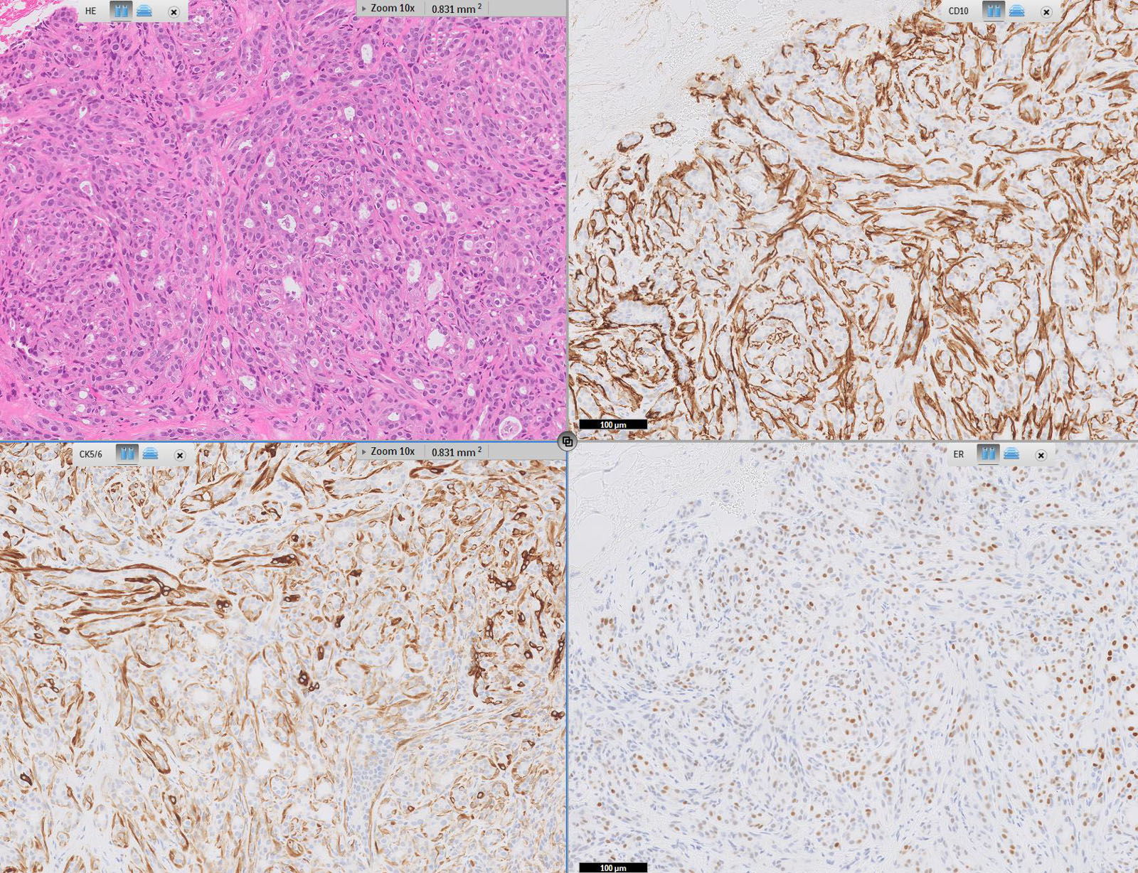


Pathology Outlines Cytokeratin 5 6 And Ck5


Pdf Triple Negative And Basal Like Breast Cancer In East Africa Zahir Moloo And Shahin Sayed Academia Edu
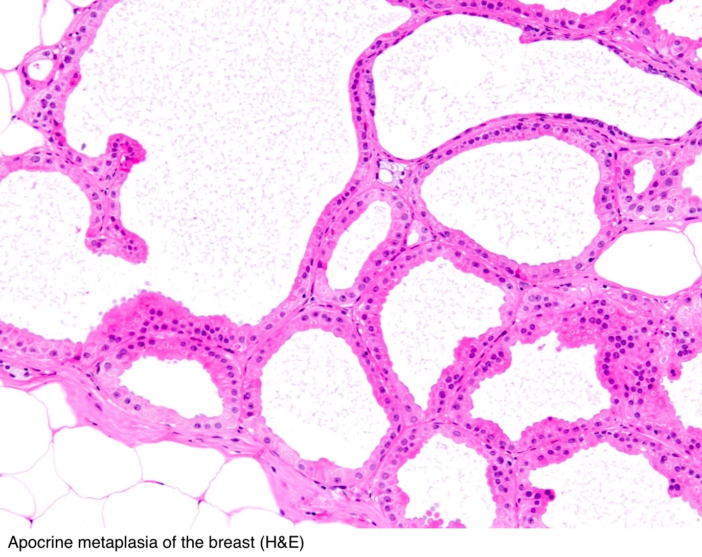


Pathology Outlines Cytokeratin 5 6 And Ck5


Expression Of P63 And Ck5 6 In Early Stage Lung Squamous Cell Carcinoma Is Not Only An Early Diagnostic Indicator But Also Correlates With A Good Prognosis Ma 15 Thoracic Cancer
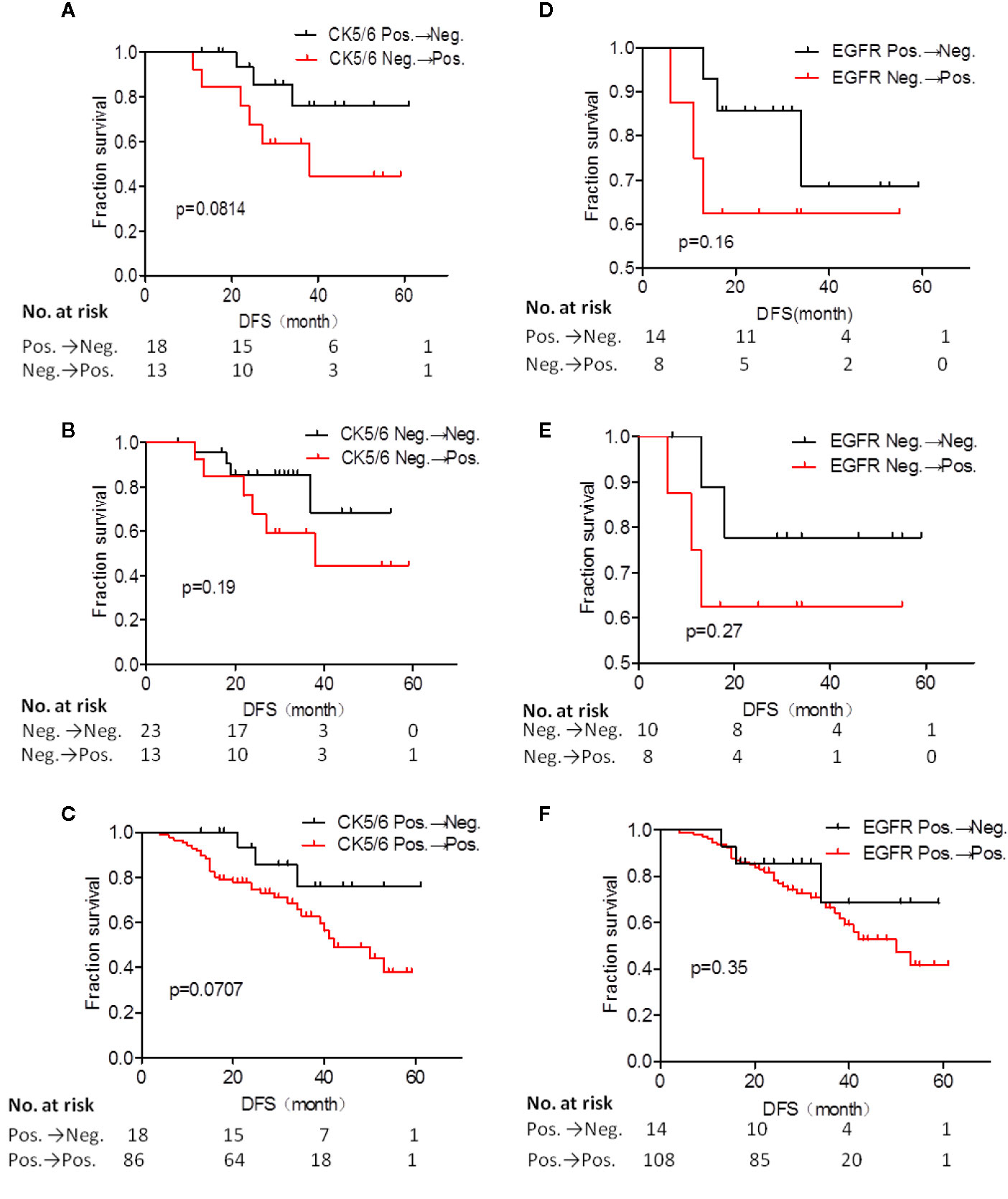


Frontiers Analysis Of Ck5 6 And Egfr And Its Effect On Prognosis Of Triple Negative Breast Cancer Oncology
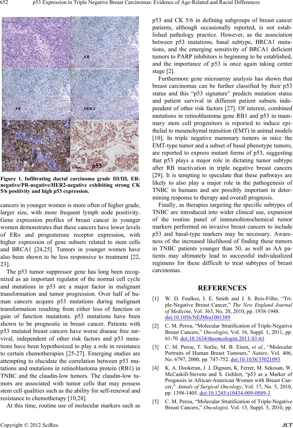


P53 Expression In Triple Negative Breast Carcinomas Evidence Of Age Related And Racial Differences


Hybrid Model Integrating Immunohistochemistry And Expression Profiling For The Classification Of Carcinomas Of Unknown Primary Site The Journal Of Molecular Diagnostics


Unclineberger Org Peroulab Wp Content Uploads Sites 1008 19 06 Aug 15 Basal Like By Ihc Nielsen Ccr 04 Pdf


Pdf Prognostic Impact Of Egfr And Ck 5 6 As Basal Markers In Triple Negative Breast Cancers Semantic Scholar
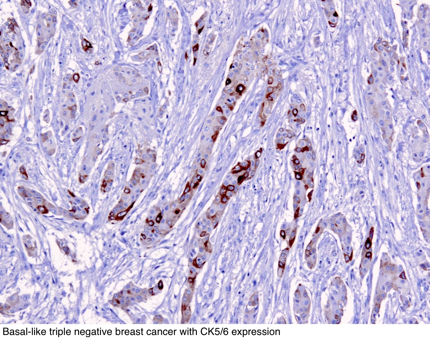


Pathology Outlines Cytokeratin 5 6 And Ck5


Prognostic Impact Of Egfr And Cytokeratin 5 6 Immunohistochemical Expression In Triple Negative Breast Cancer Sciencedirect
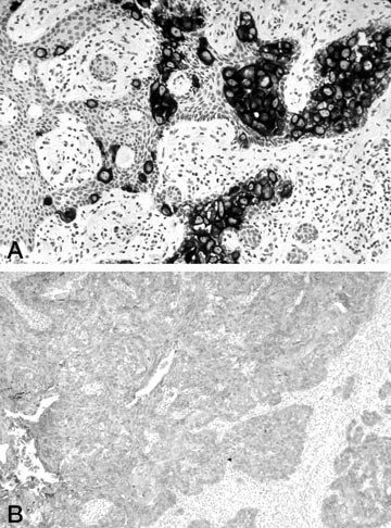


Cytokeratin 7 And Cytokeratin Expression In Epithelial Neoplasms A Survey Of 435 Cases Modern Pathology


Triple Negative Breast Carcinoma A Comparative Study Between Breast Lesion And Lymph Node Metastases A Preliminary Study


Prognostic Impact Of Egfr And Cytokeratin 5 6 Immunohistochemical Expression In Triple Negative Breast Cancer Sciencedirect


Www Pacificejournals Com Journal Index Php Apalm Article Download Apalm266 Pdf 104


Loss Of P63 And Cytokeratin 5 6 Expression Is Associated With More Aggressive Tumors In Endometrial Carcinoma Patients Stefansson 06 International Journal Of Cancer Wiley Online Library


0 件のコメント:
コメントを投稿